
First-Trimester Ultrasound Exam Guide
Published
First-Trimester Ultrasound: A Structured Exam Review
First-trimester questions test more than whether a sac is visible. They ask you to distinguish location from viability, select the right dating measurement, document early anatomy, and recognize when a finding is uncertain rather than diagnostic. This guide is for sonography education, not individualized pregnancy advice. Clinical decisions require the interpreting clinician, the complete history, and current local protocols.
Use /blog/obgyn-ultrasound-boards-study-plan for a full OB/GYN study calendar and /specialty/ob to practice case-based interpretation.
Scan Systematically: Uterus, Adnexa, and Cul-de-Sac
Establish the scan approach and orientation first. Transabdominal imaging offers a broad pelvic overview; transvaginal imaging can resolve small early structures in greater detail when appropriate and consented to. Document the uterus and endometrium, any intrauterine gestational sac, yolk sac, embryo and cardiac activity if present, both adnexa, and free fluid in the cul-de-sac. Record relevant images and measurements in the correct plane. An empty uterus on a single early study does not alone establish ectopic pregnancy or pregnancy loss.
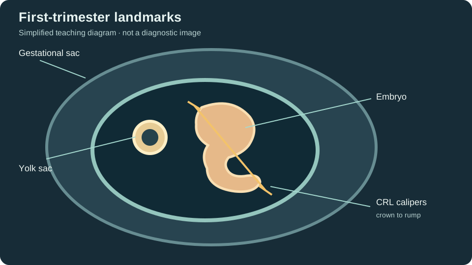
Distinguish the intrauterine sac from a nonspecific intrauterine fluid collection: a yolk sac or embryo within an intrauterine sac is stronger evidence of an intrauterine pregnancy. Examine the adnexa even when an intrauterine pregnancy is seen if symptoms or risk factors raise concern; heterotopic pregnancy is uncommon but possible, particularly after assisted reproduction. A suspected ectopic pregnancy or significant free fluid needs timely clinical assessment.
Dating: CRL Before Later Biometry
When an embryo can be measured accurately in the first trimester, crown-rump length (CRL) is the preferred ultrasound measurement for dating. Obtain a true mid-sagittal image, neutral embryo position, and calipers at crown and rump, excluding the yolk sac and limbs. If there is no measurable embryo, mean sac diameter (MSD) averages three orthogonal internal sac measurements; it is less precise for dating and must not be treated as interchangeable with CRL.
ACOG's dating guidance gives trimester- and gestational-age-specific rules for reconciling ultrasound dates with a reliable last menstrual period. A board stem may ask whether to revise an estimated due date; look at how far the two estimates differ and which gestational-age range applies. Do not memorize one universal redating threshold for every week of pregnancy.
Viability: Know What Is Diagnostic and What Needs Follow-Up
The SRU consensus uses conservative criteria to avoid a false diagnosis of pregnancy failure. Examples diagnostic of failure on transvaginal ultrasound include CRL of at least 7 mm without cardiac activity, or MSD of at least 25 mm without an embryo. Smaller measurements or findings seen before the required follow-up interval are suspicious, not definitive. The interval depends on what was visible on the earlier scan; a board question that omits the prior scan date cannot safely use an interval rule.
Common Mistake: Treating an early empty sac or a single beta-hCG value as proof of nonviability or ectopic pregnancy. Pregnancy of unknown location is a temporary description after a positive test when ultrasound has not yet established location. Serial assessment and the clinical picture matter; a so-called discriminatory beta-hCG level is not a stand-alone rule for intervention.
The presence of cardiac activity is documented, not inferred from size alone. Follow the examination protocol and avoid unnecessary exposure: the AIUM recommends prudent use of ultrasound output, particularly in early pregnancy, and M-mode rather than spectral Doppler is generally preferred when documenting early embryonic heart activity.
Early Anatomy and Screening Are Different Tasks
A first-trimester survey documents visible structures appropriate to gestational age, such as the embryo/fetus, number of fetuses, chorionicity when multiple gestations are present, and early cranial, abdominal, and limb anatomy when assessable. A normal early examination does not replace the detailed second-trimester anatomic survey. Image quality and what can be evaluated vary with gestational age, fetal position, equipment, and patient factors.
Nuchal translucency is a specific screening measurement made within an established gestational-age and CRL window by trained operators; it is not a general neck-fold observation and does not by itself diagnose a chromosome condition. Follow the applicable screening program's technique and interpretation rules rather than extrapolating a threshold from a still image. For later fetal assessment, see /blog/fetal-echocardiography-boards-high-yield.
Common Board-Question Pitfalls
Location versus viability. A correctly located pregnancy may not yet have demonstrable cardiac activity; a visible sac-like structure alone does not settle location. State which question the image answers.
Measurement choice. Use CRL when a measurable embryo is present, not MSD or second-trimester BPD. Make sure calipers include the correct endpoints and the image is not oblique.
Certainty of language. Separate findings diagnostic of failure from those suspicious for it. Look for precise CRL/MSD measurements and the timing and contents of an earlier scan.
Safety and limitations. Doppler settings chosen for a fetal vessel later in pregnancy are not automatically appropriate for a tiny early embryo. An early normal survey cannot exclude every anomaly.
To review related imaging physics, use /blog/ultrasound-artifacts-visual-guide; refraction can produce a duplicated sac-like image. For focused OB study materials visit /specialty/ob.
Frequently Asked Questions
Q: What is the best first-trimester measurement for dating?
A technically adequate crown-rump length when an embryo is measurable. MSD is less precise and is not a substitute when CRL is available.
Q: Does an empty uterus with a positive pregnancy test mean ectopic pregnancy?
No. It may represent a very early intrauterine pregnancy, an early loss, or an ectopic pregnancy. The evaluation includes adnexa, free fluid, clinical assessment, and appropriate follow-up.
Q: Can I diagnose pregnancy failure with a CRL under 7 mm and no heartbeat?
Not on that measurement alone. The SRU diagnostic cutoff is CRL at least 7 mm without cardiac activity on transvaginal imaging; smaller embryos require appropriate follow-up.
Q: Does a normal early scan replace the mid-trimester anatomy survey?
No. Some anatomy is not yet assessable or is limited by the imaging conditions. The later detailed survey remains important.
Study Takeaway
Organize every first-trimester stem around location, dating, viability, and anatomy. Check how the image was acquired, measure only what can be measured reliably, and avoid promoting a suspicious finding into a definitive diagnosis. Pair these concepts with /blog/obgyn-ultrasound-boards-study-plan and timed cases at /specialty/ob.
Sources & Further Reading
- Diagnostic Criteria for Nonviable Pregnancy Early in the First Trimester (SRU consensus) — New England Journal of Medicine / SRU
- Methods for Estimating the Due Date — ACOG
- AIUM Official Statements and Practice Parameters — AIUM
- Obstetrics and Gynecology examination — Inteleos / ARDMS
If you find this article helpful and want to put the strategies into practice, sign up for an Ultrasound Analytics account to access the full ARDMS-aligned question bank, AI tutoring on every missed answer, full-length timed exams, and the analytics dashboard that translates your performance into a Readiness Score for each specialty registration.